
Unveiling the Cell Membrane and its Transport Mechanisms
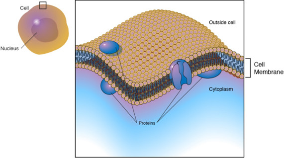 Cell Membrane Structure
The cell membrane, often referred to as the plasma membrane, serves as the vital outer boundary of every cell. This intricate lipid bilayer meticulously separates the cell’s internal components from the external environment. Acting as a selective gatekeeper, the cell membrane dictates which substances can pass in and out, ensuring the cell’s survival and proper function. Understanding Transport Across Cell Membranes is fundamental to grasping cellular biology and physiology.
Delving into the Composition of the Cell Membrane
The cell membrane’s sophisticated functionality stems from its unique composition. As illustrated in Figure 1, it’s not merely a simple barrier but a dynamic assembly of several key components:
- Phospholipid Bilayer: This is the foundational structure, featuring hydrophilic (polar) heads facing outwards, interacting with the watery environments both inside and outside the cell. Conversely, hydrophobic (nonpolar) fatty acid tails are tucked inwards, forming the membrane’s core. The presence of unsaturated fatty acids within these tails enhances membrane fluidity – a crucial characteristic for membrane function. A higher proportion of unsaturated fatty acids equates to greater fluidity.
- Cholesterol: Interspersed within the phospholipid bilayer, cholesterol molecules play a vital role in modulating membrane rigidity. Increased cholesterol content leads to a more rigid and stable membrane structure.
- Proteins: Two primary types of proteins are embedded within or attached to the cell membrane:
- Integral Proteins: These proteins are deeply embedded within the membrane, often spanning its entire width. They primarily function as transporters, facilitating the movement of specific molecules and ions across the membrane.
- Peripheral Proteins: Loosely associated with the membrane’s outer surface, these proteins often act as enzymes, catalyzing reactions that occur at the membrane surface and mediating interactions with the cell’s surroundings.
- Glycoproteins and Glycolipids: These components feature carbohydrate chains attached to either proteins (glycoproteins) or lipids (glycolipids) on the cell’s exterior surface. They are crucial for cell-to-cell recognition, enabling cells to identify and interact with each other.
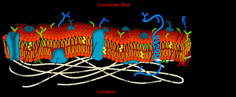 Detailed Cell Membrane Diagram
Figure 1: A detailed illustration of the cell membrane structure, highlighting the phospholipid bilayer, cholesterol, integral and peripheral proteins, and glycoproteins/glycolipids.
Beyond controlling the traffic of molecules, the cell membrane is also involved in cell adhesion, cell signaling, and providing a framework for the cytoskeleton. The cytoskeleton, an internal network of protein fibers, helps maintain cell shape and anchors the cell to the extracellular matrix, contributing to tissue integrity.
Mechanisms of Transport Across the Cell Membrane
To sustain life, cells must selectively exchange substances with their environment. The cell membrane facilitates this exchange through various transport mechanisms, allowing for the import of essential nutrients and the export of waste products. These mechanisms can be broadly categorized based on their energy requirements and the nature of molecules they transport.
Diffusion: Passive Movement Down the Concentration Gradient
Diffusion is a fundamental process of passive transport, meaning it doesn’t require the cell to expend energy. It describes the movement of molecules from an area of higher concentration to an area of lower concentration, driven by the inherent kinetic energy of molecules and the tendency to reach equilibrium.
Small, nonpolar molecules, such as oxygen (O2), carbon dioxide (CO2), and nitrogen (N2), readily diffuse across the cell membrane. Their size and nonpolar nature allow them to pass through the hydrophobic core of the lipid bilayer without assistance. This process is crucial for gas exchange in respiration and photosynthesis.
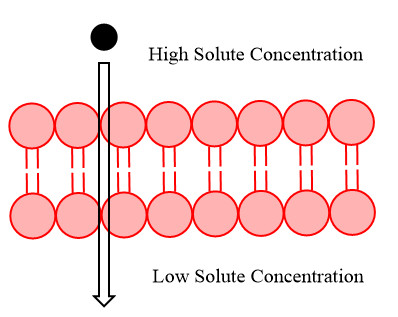 Diffusion Across Cell Membrane
Figure 2: Illustration depicting simple diffusion of molecules across the cell membrane from a high concentration area to a low concentration area.
Facilitated Diffusion: Protein-Assisted Passive Transport
While diffusion works well for small, nonpolar molecules, the cell membrane presents a barrier to larger, polar, or charged substances. Facilitated diffusion provides a pathway for these molecules to cross the membrane passively, aided by membrane proteins. This process is still considered passive transport because it relies on the concentration gradient and does not require cellular energy expenditure.
Membrane proteins involved in facilitated diffusion can be either channel proteins or carrier proteins. Channel proteins form hydrophilic pores or channels through the membrane, allowing specific ions or small polar molecules to pass through. Carrier proteins, on the other hand, bind to the molecule to be transported, undergo a conformational change, and release the molecule on the other side of the membrane.
Examples of molecules transported via facilitated diffusion include ions like chloride (Cl⁻) and bicarbonate (HCO₃⁻), and larger polar molecules such as glucose. Water, despite being polar, also utilizes facilitated diffusion through specialized channel proteins called aquaporins, facilitating rapid water movement across membranes, which is essential for osmosis and maintaining cell volume.
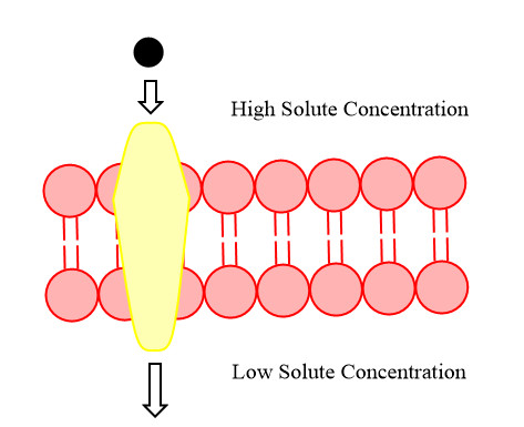 Facilitated Diffusion with Protein Channels
Figure 3: Diagram illustrating facilitated diffusion, where membrane proteins assist in the transport of molecules across the cell membrane, following the concentration gradient.
Active Transport: Energy-Driven Movement Against the Gradient
In contrast to passive transport, active transport enables cells to move substances against their concentration gradient – from an area of lower concentration to an area of higher concentration. This “uphill” movement requires energy input, typically in the form of ATP (adenosine triphosphate), the cell’s energy currency. Active transport is crucial for maintaining cellular homeostasis and establishing concentration gradients essential for various cellular processes.
Active transport is always mediated by membrane proteins, often referred to as pumps. These protein pumps utilize the energy from ATP hydrolysis to actively transport specific ions or molecules across the membrane. A prime example is the sodium-potassium pump, which actively transports sodium ions (Na⁺) out of the cell and potassium ions (K⁺) into the cell. This pump is vital for maintaining the electrochemical gradient across nerve and muscle cell membranes, essential for nerve impulse transmission and muscle contraction.
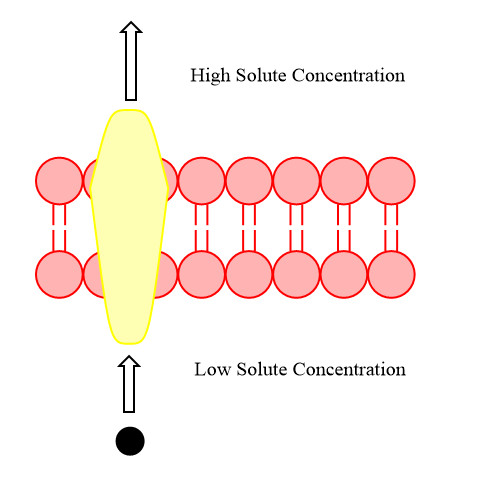 Active Transport Against Concentration Gradient
Figure 4: Illustration of active transport, showing the movement of molecules against the concentration gradient, requiring energy input, and facilitated by protein pumps.
The Electron Transport Chain: An Example of Facilitated Diffusion and Active Transport in Concert
The electron transport chain (ETC), a critical component of cellular respiration, beautifully exemplifies both facilitated diffusion and active transport working in tandem. Located within the inner mitochondrial membrane, the ETC is responsible for generating the majority of ATP in eukaryotic cells.
Following the citric acid cycle, energy-rich molecules NADH and FADH2 are produced. The ETC harnesses the energy stored in these molecules to generate ATP. The ETC comprises a series of protein complexes embedded in the inner mitochondrial membrane. Electrons from NADH and FADH2 are passed along these complexes in a sequential manner, losing small amounts of energy at each step.
This energy released during electron transfer is utilized to actively pump protons (H⁺ ions) from the mitochondrial matrix (low H⁺ concentration) to the intermembrane space (high H⁺ concentration). This active transport of protons creates an electrochemical gradient across the inner mitochondrial membrane, with a higher concentration of H⁺ in the intermembrane space.
Subsequently, protons flow down this electrochemical gradient, from the intermembrane space back into the matrix, through a specific channel protein called ATP synthase. This flow of protons is an example of facilitated diffusion. ATP synthase harnesses the energy released during this facilitated diffusion of protons to convert ADP (adenosine diphosphate) and inorganic phosphate into ATP, a process termed oxidative phosphorylation.
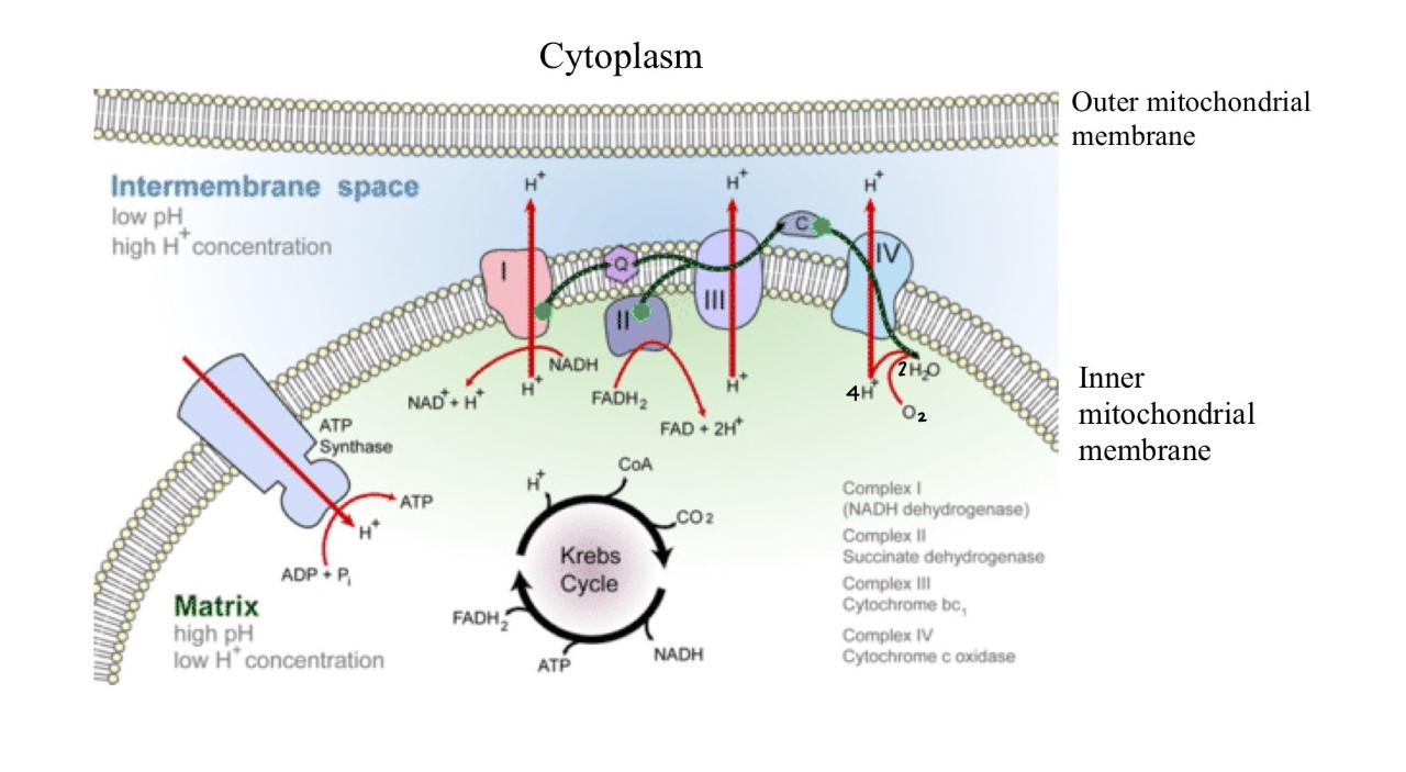 Electron Transport Chain in Mitochondria
Figure 5: A schematic representation of the electron transport chain in the inner mitochondrial membrane, illustrating the active transport of protons and facilitated diffusion of protons through ATP synthase for ATP production.
Oxygen acts as the final electron acceptor in the ETC, combining with electrons and protons to form water. Through glycolysis, the citric acid cycle, and the electron transport chain, a single glucose molecule can yield approximately 32 ATP molecules. Cellular respiration effectively converts the chemical energy stored in glucose into readily usable energy in the form of ATP, powering various cellular activities, including active transport processes and biosynthesis of essential biomolecules.
Conclusion: The Dynamic Nature of Membrane Transport
In summary, transport across the cell membrane is a multifaceted process vital for cellular life. Cells employ three primary mechanisms – diffusion, facilitated diffusion, and active transport – to selectively move substances across their membranes. Diffusion and facilitated diffusion are passive processes driven by concentration gradients, while active transport requires energy to move substances against their gradients. Understanding these transport mechanisms is crucial for comprehending cellular function, energy production, and the intricate interplay between cells and their environment.